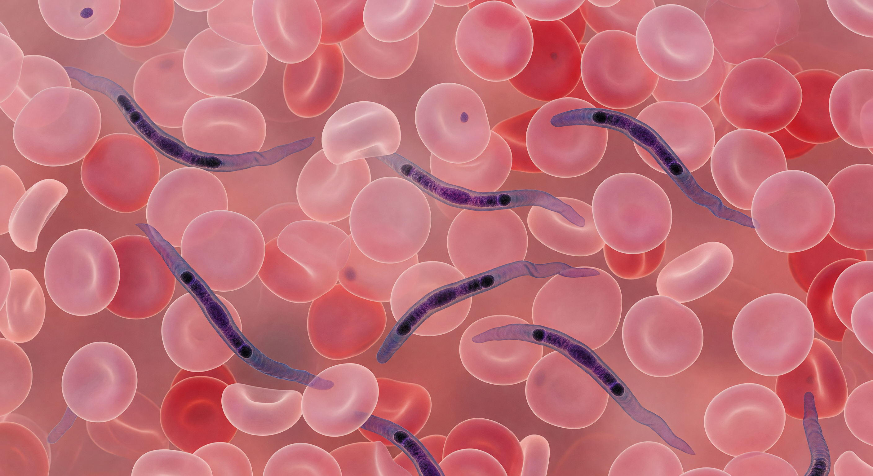
The view is an overwhelming immersion inside living tissue — in every direction, soft rose-pink biconcave discs press close, their thinned centers transmitting amber light as faint aureoles, their thickened carmine rims overlapping until the visual field dissolves into a blush fog of stacked geometry with no horizon and no escape. Between these densely crowded erythrocytes, each approximately seven micrometres across, Trypanosoma brucei navigate the narrow interstices like sinuous calligraphy — elongated blue-violet bodies fifteen to thirty micrometres long, their undulating flagellar membranes propagating slow silk-ribbon waves that represent the parasite's primary means of locomotion through a fluid medium governed entirely by viscous drag rather than inertia. Giemsa staining saturates the scene with its characteristic palette, concentrating in the dense chromatin of the nucleus and the compact kinetoplast — that extraordinary mitochondrial DNA mass unique to kinetoplastid protists, sitting near the posterior end of each parasite body like a dark seed, its position marking the base of the flagellum and anchoring the entire motility apparatus. These organisms are not incidental passengers but active architects of pathology: Trypanosoma brucei crosses the blood-brain barrier to cause African sleeping sickness, and the undulating movement visible here is the same swimming behavior that allows the parasite to evade host immune surveillance while traveling through exactly this claustrophobic corridor of red cells and plasma.
Other languages
- Français: Trypanosomes Serpentant Entre Globules
- Español: Tripanosomas Serpenteando Entre Glóbulos
- Português: Tripanossomos Serpenteando Entre Hemácias
- Deutsch: Trypanosomen Schlängeln Zwischen Blutzellen
- العربية: طفيليات المثقبيات بين خلايا الدم
- हिन्दी: लाल कोशिकाओं के बीच ट्रिपैनोसोमा सर्प
- 日本語: 赤血球の間を泳ぐトリパノソーマ
- 한국어: 적혈구 사이 트리파노소마 기생충
- Italiano: Tripanosomi Serpeggianti Tra Globuli Rossi
- Nederlands: Trypanosomen Kronkelen Tussen Rode Cellen