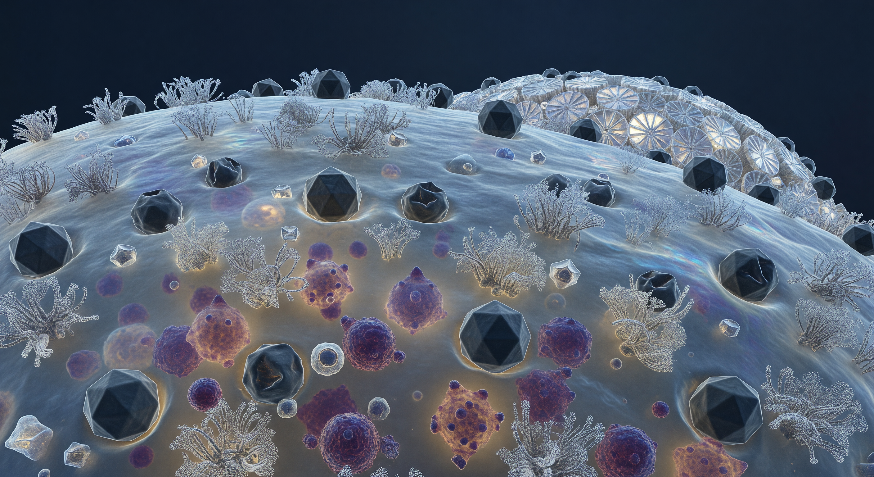
The viewer hangs motionless above a gently curving biological horizon, close enough that the coccolithophore's outer membrane resolves into a trembling, semi-translucent skin — a landscape of glycoprotein clusters rising like coral formations from a blue-gray surface shimmering with thin-film interference where lipid raft domains concentrate. Pressed into this terrain at dozens of sites, dark icosahedral capsids of *Emiliania huxleyi* virus sit in various states of predatory engagement: some geometrically pristine, their twenty facets catching cold diffuse light along sharp ridges, others visibly collapsed inward — capsid shells crumpled like punctured geodesic domes after successful genome injection, the membrane dimpled beneath each one in a shallow hollow of biological surrender. Through the translucent membrane wall, the cytoplasm glows like fogged amber glass, crowded with dense irregular masses of deep purple and magenta — viral progeny assembling by the hundreds on nucleoprotein scaffolds in a factory-scale replication event that presses outward against the cell wall from below, bulging the membrane like overfilled sacking. Along the cell's sweeping curvature toward the distant coccosphere horizon, interlocking calcite coccolith plates rise as white ceramic buttresses, their spoke-and-rim geometry scattering what little deep-water light reaches them into brief prismatic flashes — a world of extraordinary crystalline precision and molecular beauty quietly unraveling, every contested surface a boundary between the cell's remaining chemistry and the viral program steadily overwriting it.
Other languages
- Français: Siège Viral sur Cellule
- Español: Asedio Viral en Célula
- Português: Cerco Viral na Célula
- Deutsch: Viraler Angriff auf Zelle
- العربية: حصار فيروسي للخلية
- हिन्दी: कोशिका पर वायरल घेरा
- 日本語: 細胞表面のウイルス包囲
- 한국어: 세포 표면 바이러스 포위
- Italiano: Assedio Virale Cellulare
- Nederlands: Virale Belegering Celoppervlak