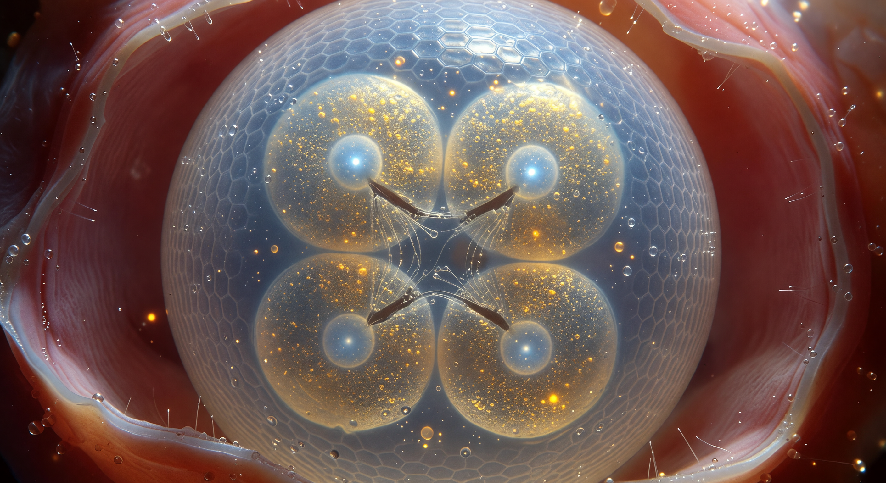
You hover so close to the eggshell that its curved face fills your entire field of view like the surface of a pale, frost-lit moon — a compressed chitinous lattice of interlocking polysaccharide fibers that scatters polarized light into shifting bands of indigo and silver, cool and luminous against the warm rose darkness of the surrounding uterine tissue. Behind this translucent barrier, four blastomeres press against one another in tight tetrahedral geometry, their flattened contact surfaces forming sharp, dark cleavage furrows while their interiors glow with dense golden yolk granules — autofluorescent masses of lipid and protein reserves laid down to fuel every division to come, from this first quartet of cells to the precisely invariant 959 somatic cells of the adult. At the center of each blastomere, a pale arctic-blue nucleus holds a single brilliant nucleolus, the ribosome-assembly engine running at maximum output for the demands of rapid embryogenesis, while between two cells the fading silver threads of a mitotic spindle — microtubules just completing chromosome segregation — catch the ambient light in brief metallic gleams before depolymerizing back into cytoplasmic tubulin pools. The perivitelline fluid sealing the space between embryo and shell is optically perfect, a pressurized aqueous medium that maintains the egg's shape while providing a chemically buffered, mechanically cushioned world in which this ancient, conserved developmental program — unchanged in its essential logic across hundreds of millions of years — executes with clockwork precision.
Other languages
- Français: Intérieur Doré Embryon Quatre Cellules
- Español: Interior Dorado Embrión Cuatro Células
- Português: Interior Dourado Embrião Quatro Células
- Deutsch: Vierzelliger Embryo Goldenes Innere
- العربية: داخل الجنين الذهبي الرباعي
- हिन्दी: चार-कोशिका भ्रूण स्वर्णिम अंतर
- 日本語: 四細胞胚の黄金の内部
- 한국어: 사세포 배아 황금 내부
- Italiano: Embrione Quattro Cellule Interno Dorato
- Nederlands: Viercellig Embryo Gouden Interieur