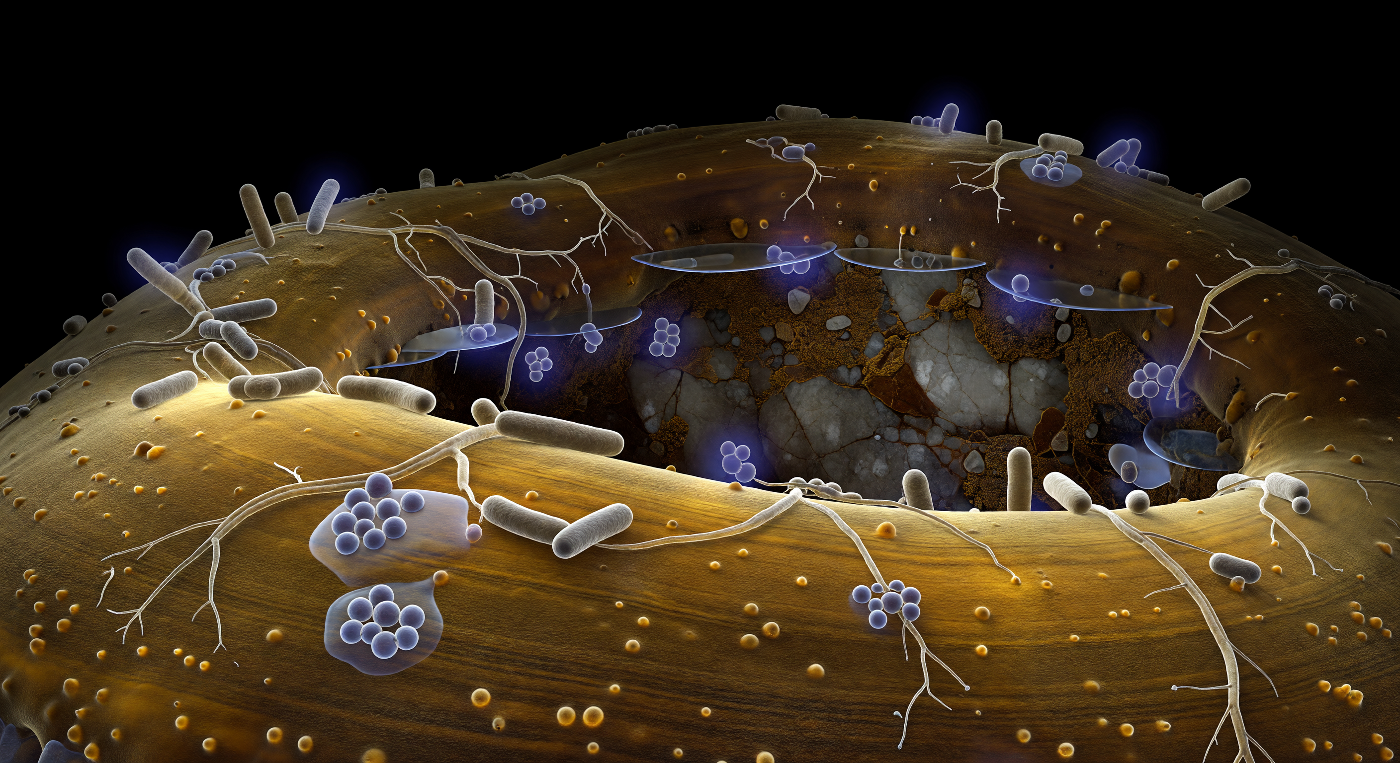
The curved amber wall rising before you is the outer surface of an ectomycorrhizal hypha — a single fungal filament perhaps seven micrometers wide, yet reading here as a glazed sandstone escarpment that fills your entire horizon, its chitinous laminations glowing from within as cold cytoplasmic light filters through semi-translucent layers of polysaccharide and glycoprotein. Across this hillside topography, rod-shaped bacteria of the genus *Bacillus* and branching *Streptomyces* filaments constitute a hyphosphere microbiome — mycorrhiza helper bacteria known to suppress pathogens, solubilize nutrients, and produce phytohormones that enhance fungal colonization of host roots, forming one of the most ecologically significant microbial guilds in forest soil. The bacteria cling and sprawl in small waxy colonies bridged by exopolysaccharide gels that pool between cells like spilled glycerin, anchoring the community to the hyphal wall through adhesive tethers visible only where the cytoplasmic glow catches their contact angle; individuals broadcasting chemical signals into the surrounding aqueous film appear wreathed in faint violet diffusion halos, their molecular messages dissolving within a few cell-lengths into the water that fills this subterranean pore. Beyond the hypha's curvature, a cavernous soil void opens into absolute darkness, its far wall a fractured feldspar face surfaced with organic films the color of dark toffee, bridged to the hypha by mirror-flat water menisci that catch the amber glow in single glinting reflections. There is no light from above — only chemistry, membrane boundaries, and the permanent collaborative negotiation of living surfaces pressed against one another in the lightless architecture beneath the forest floor.
Other languages
- Français: Surface Microbienne de l'Hyphosphère
- Español: Superficie Bacteriana de la Hifosfera
- Português: Superfície Bacteriana da Hifosfera
- Deutsch: Hyphosphären Bakterien Mikrokolonie
- العربية: سطح مستعمرات بكتيريا الهيفوسفير
- हिन्दी: हाइफोस्फीयर जीवाणु सूक्ष्म-उपनिवेश
- 日本語: 菌糸圏細菌マイクロコロニー表面
- 한국어: 균사권 세균 미세집락 표면
- Italiano: Superficie Batterica dell'Ifosfera
- Nederlands: Hyfosfeerbacteriën Microkolonie Oppervlak