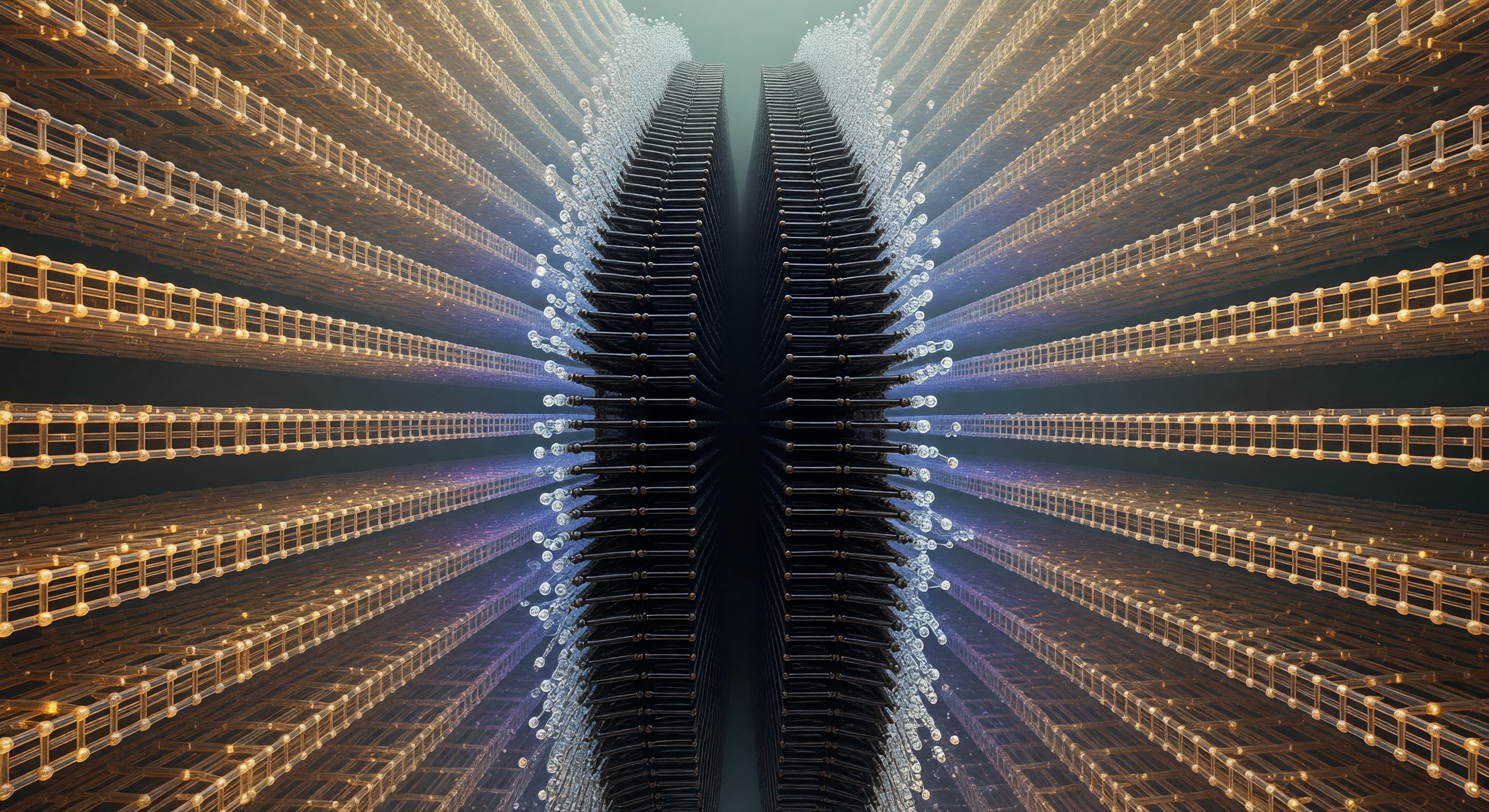
Looking straight down the axis of an amyloid-β fibril, the viewer enters an architectural corridor of almost hallucinatory regularity — parallel β-sheet layers stacked with sub-nanometer precision recede into apparent infinity, each plane of peptide backbone an exact structural echo of the one before it, separated by intervals of roughly 4.7 ångströms governed entirely by hydrogen-bond geometry rather than any external template. Those hydrogen bonds themselves appear as luminous amber-gold rungs spanning laterally between adjacent strands, their directionality perpendicular to the recession axis, tracing a ladder whose terminus is never reached — each N–H donor locked to a carbonyl oxygen acceptor with a bond distance near 2 Å, the quantum mechanical overlap of molecular orbitals rendered as warm, continuous luminescence. At the absolute center of the abyss lies the steric-zipper core, a seam of profound darkness where nonpolar side chains — leucines, isoleucines, valines — interdigitate across the protofilament interface with van der Waals contact so complete that bulk water is entirely excluded, the two apposed β-sheet faces held together by London dispersion forces across a boundary that is, at the molecular level, a seamless coalescence of complementary hydrophobic surfaces. Flanking this dry obsidian corridor, the outer fibril surface shimmers with a blue-white corona of ordered solvation water, each molecule oriented by the polar backbone beneath, hydrogen-bond lifetimes of one to ten picoseconds creating a nacreous thermal shimmer at the liquid-ordered interface. The overwhelming sensation is one of self-templating inevitability: this structure assembled itself one molecular layer at a time, each incoming peptide recognizing and replicating the cross-β geometry of its predecessor with a fidelity that feels less like biology than like crystallographic law made animate.
Other languages
- Français: Abîsse Bêta des Fibrilles
- Español: Abismo Fibrilar Amiloide
- Português: Abismo Fibrilar Amiloide
- Deutsch: Amyloid Fibril Kreuzabgrund
- العربية: هاوية ألياف النشواني
- हिन्दी: एमिलॉयड फाइब्रिल गहन खाई
- 日本語: アミロイド繊維の深淵
- 한국어: 아밀로이드 피브릴 심연
- Italiano: Abisso Fibrillare Amiloide
- Nederlands: Amyloïde Fibril Bèta Afgrond