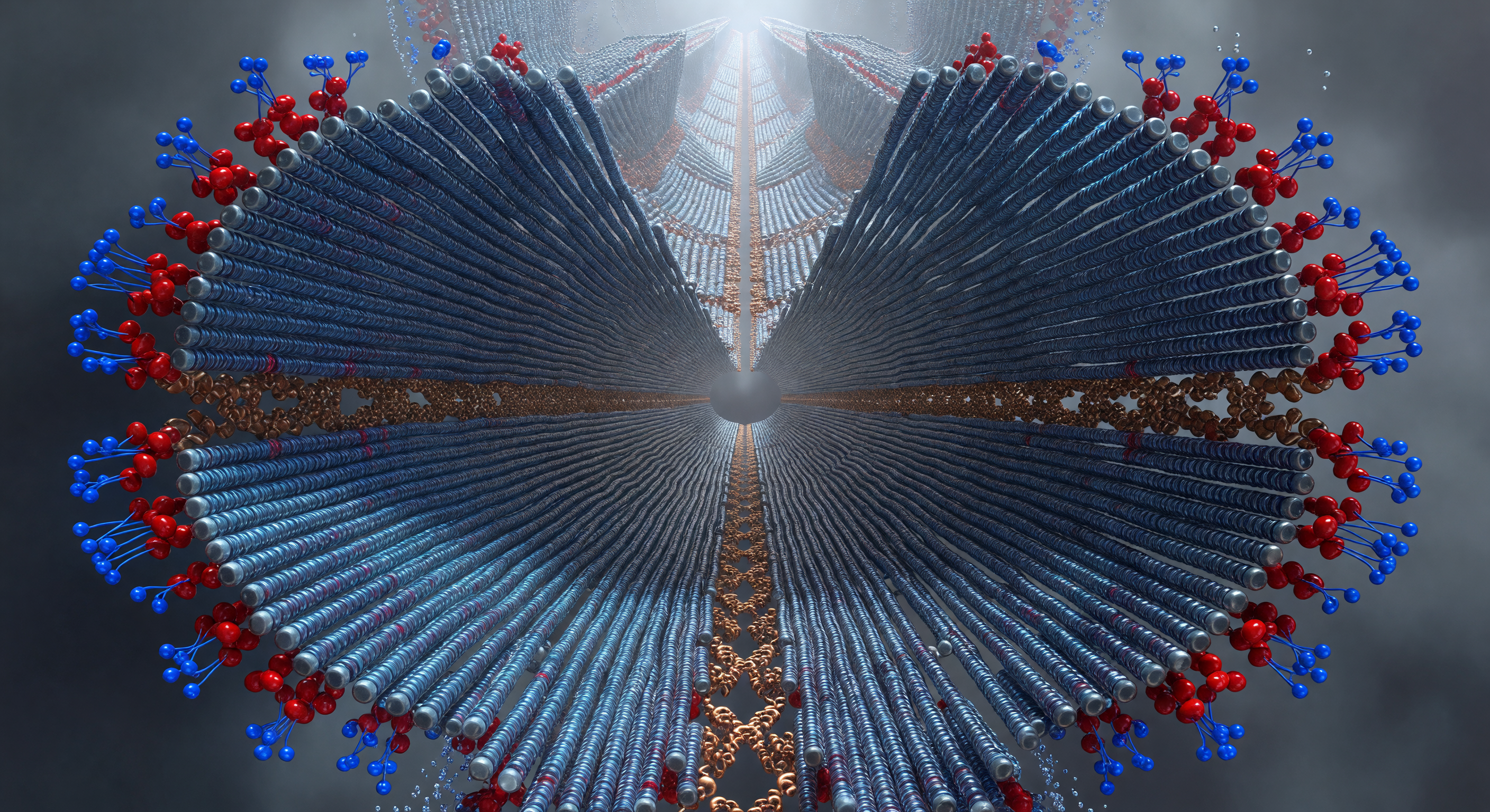
You are suspended at the dead center of the fibril axis, looking down a tunnel of four protofilament lobes that sweep outward from you like the petals of a gothic flower, each wing a curved wall of beta-strands stacked in relentless 4.7-ångström increments — a spacing so precise it functions less as a measurement than as the structural heartbeat of an entire category of molecular pathology. These are amyloid fibrils: cross-beta assemblies formed when once-soluble proteins collapse out of their native conformations and lock into hydrogen-bonded sheets that propagate laterally with a thermodynamic permanence rivaling crystalline mineral. At the core, between the four protofilaments, a steric zipper seals the structure from within — interdigitated side chains meshed in near-contact, van der Waals surfaces touching across a gap so narrow that bulk water is excluded entirely, replaced by a dry, amber-glowing compression of electron density that drives stability through hydrophobic burial and geometric complementarity too exact to be accidental. At the outer frontier, glutamate and lysine residues reach into the surrounding solvent haze — electrostatically charged sentinels creating the dielectric fringe that both stabilizes the assembly against lateral aggregation and presents the surface chemistry that living systems cannot easily degrade. The entire structure, roughly 10 nanometers across, extends for hundreds of nanometers along the axis receding above you: at this scale, that distance reads as geological, a molecular colonnade built from the slow catastrophe of misfolding.
Other languages
- Français: Coupe transversale fibrille amyloïde
- Español: Sección transversal fibra amiloide
- Português: Corte transversal fibra amiloide
- Deutsch: Amyloidfibril Kernquerschnitt
- العربية: مقطع نواة خيط نشواني
- हिन्दी: एमिलॉइड फाइब्रिल क्रॉस-सेक्शन
- 日本語: アミロイド線維核断面
- 한국어: 아밀로이드 섬유 핵 단면
- Italiano: Sezione trasversale fibrilla amiloide
- Nederlands: Amyloïde fibril kern dwarsdoorsnede