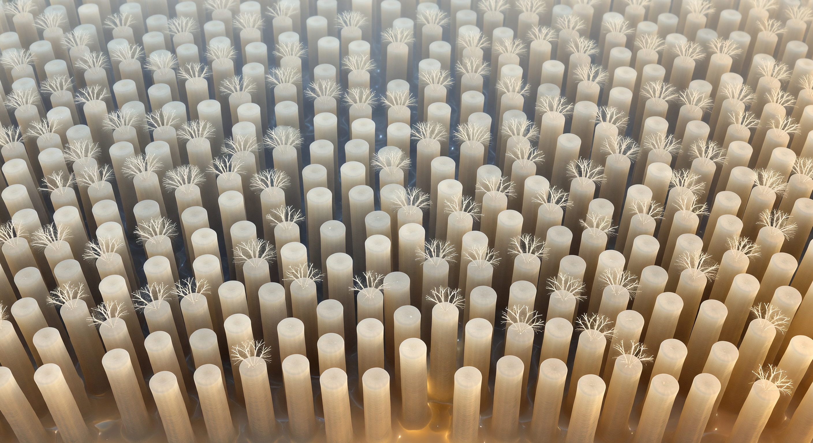
You are suspended at the very tips of the brush border, looking obliquely across a landscape that extends in every direction like an engineered plain — thousands upon thousands of cylindrical microvillus shafts, each roughly 100 nanometers across, packed into near-hexagonal regularity and rising to meet you from the enterocyte surface far below. These projections are not passive scaffolding: each shaft is stiffened internally by a tightly cross-linked bundle of actin filaments anchored through villin, fimbrin, and myosin-1a into the dense terminal web of the cell cortex beneath, the entire array constituting the intestinal brush border — a structure that amplifies the absorptive surface area of the small intestine by a factor of roughly twenty. From every shaft tip rises the glycocalyx, a soft three-dimensional pelt of O- and N-linked oligosaccharide chains extending from transmembrane glycoproteins including alkaline phosphatase and the peptide hydrolases embedded in each villus membrane, their combined mass forming a continuous extracellular matrix several tens of nanometers deep that functions simultaneously as molecular sieve, enzymatic surface, and protective barrier against luminal proteases. Oblique warm light rakes across the field from the upper right, casting precise crescent shadows in the narrow intervillus channels — those aqueous gutters between shafts, rich in ions, partially hydrolyzed peptides, and monosaccharides, the very molecules that will cross the membrane below through SGLT1 and GLUT5 transporters in the next fraction of a second. The slight tilt of one shaft, the asymmetric thickening of a glycocalyx cloud on its windward face, the faint oscillatory tension encoded in this frozen moment — all of it declares living tissue caught mid-breath, restless and continuous beneath the stillness of the image.
Other languages
- Français: Pointes de microvillosités intestinales
- Español: Puntas de microvellosidades intestinales
- Português: Pontas de microvilosidades intestinais
- Deutsch: Mikrovilli-Spitzen des Bürstensaums
- العربية: أطراف الزغيبات المعوية الدقيقة
- हिन्दी: आंत्र ब्रश बॉर्डर सूक्ष्म रोम शीर्ष
- 日本語: 腸管微絨毛の先端群
- 한국어: 장 미세융모 끝단 풍경
- Italiano: Punte di microvilli intestinali
- Nederlands: Toppen van intestinale microvilli