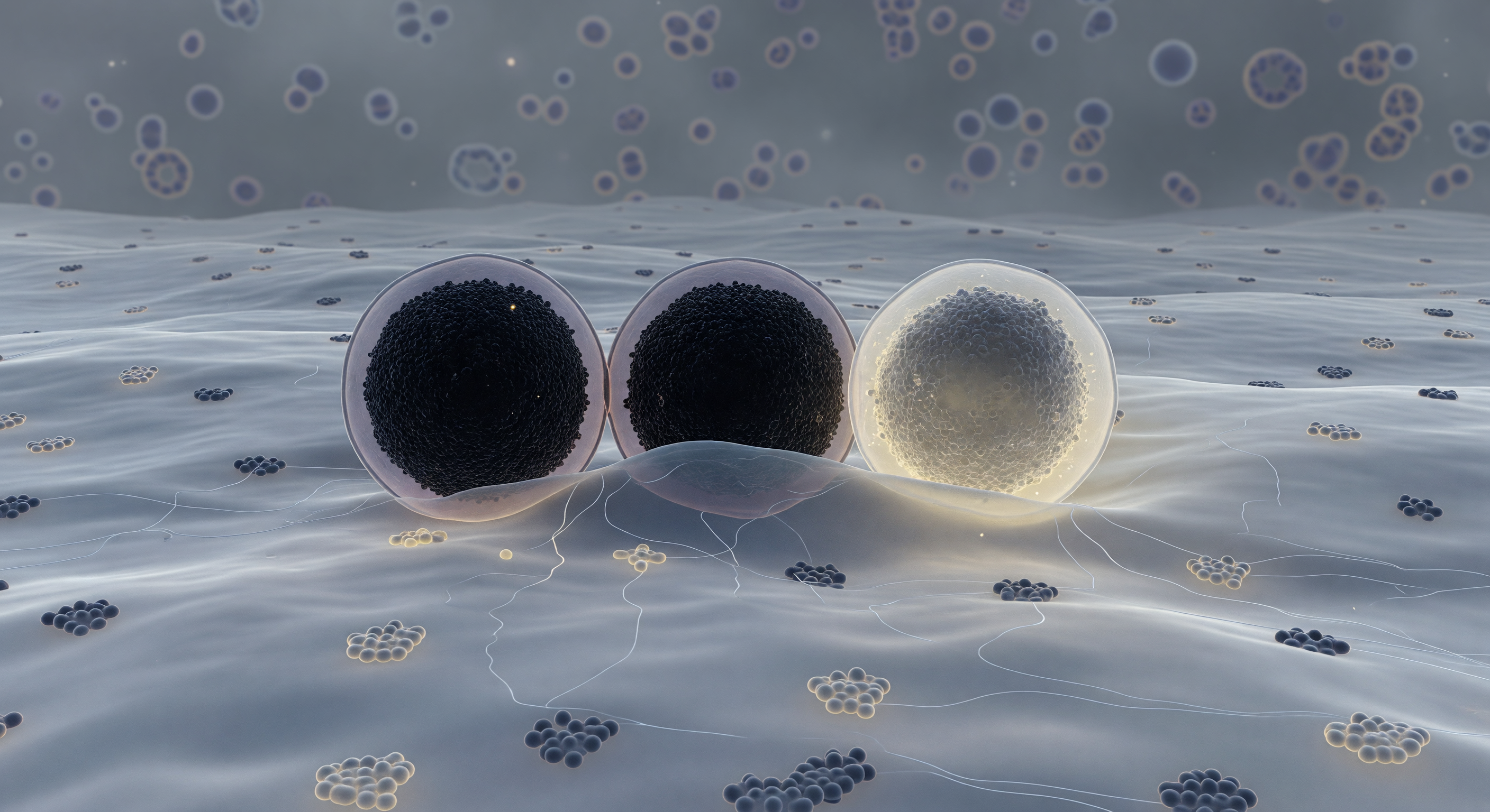
You exist pressed against the inner face of a plasma membrane that stretches to every horizon like an immense, softly luminous plain — a fluid bilayer of blue-grey lipid faintly threaded with embedded protein complexes, its surface alive with slow rolling topography where cholesterol-rich raft domains pool in subtly lighter, more ordered patches. Three granules dominate the scene, each the scale of a small building: the first sits docked in full contact, its near-black crystalline core of zinc-insulin hexamers so densely packed they absorb almost all light, ringed by a thin lavender halo where its limiting membrane holds; the second bows the plasma membrane gently inward in a hemi-fused collar where lipid identity dissolves into indeterminate grey; the third has already surrendered, its once-opaque core unraveling from deep charcoal to a dispersing warm ivory-yellow haze as insulin molecules escape into the pale amber luminosity of the extracellular space beyond. This is the final choreography of regulated exocytosis, driven by SNARE protein complexes — syntaxin, SNAP-25, and synaptobrevin — that zipper the two bilayers together with enough mechanical force to overcome the energy barrier of membrane fusion, a process completed in milliseconds yet representing the culmination of hours of granule biogenesis and priming. Threading through the cytoplasmic space between the granules, ghostly actin filaments form a loose cortical mesh, while the deeper cytoplasm recedes into a structured murk of macromolecular crowding — a single frozen instant in a thermally inevitable act, caught mid-breath, that cannot be undone.
Other languages
- Français: Zone d'ancrage des granules
- Español: Zona de anclaje de gránulos
- Português: Zona de ancoragem de grânulos
- Deutsch: Insulin-Granula-Andockzone
- العربية: منطقة إرساء حبيبات الأنسولين
- हिन्दी: इंसुलिन कणिका डॉकिंग क्षेत्र
- 日本語: インスリン顆粒ドッキング帯
- 한국어: 인슐린 과립 도킹 구역
- Italiano: Zona di ancoraggio dei granuli
- Nederlands: Insulinekorrel dockingzone