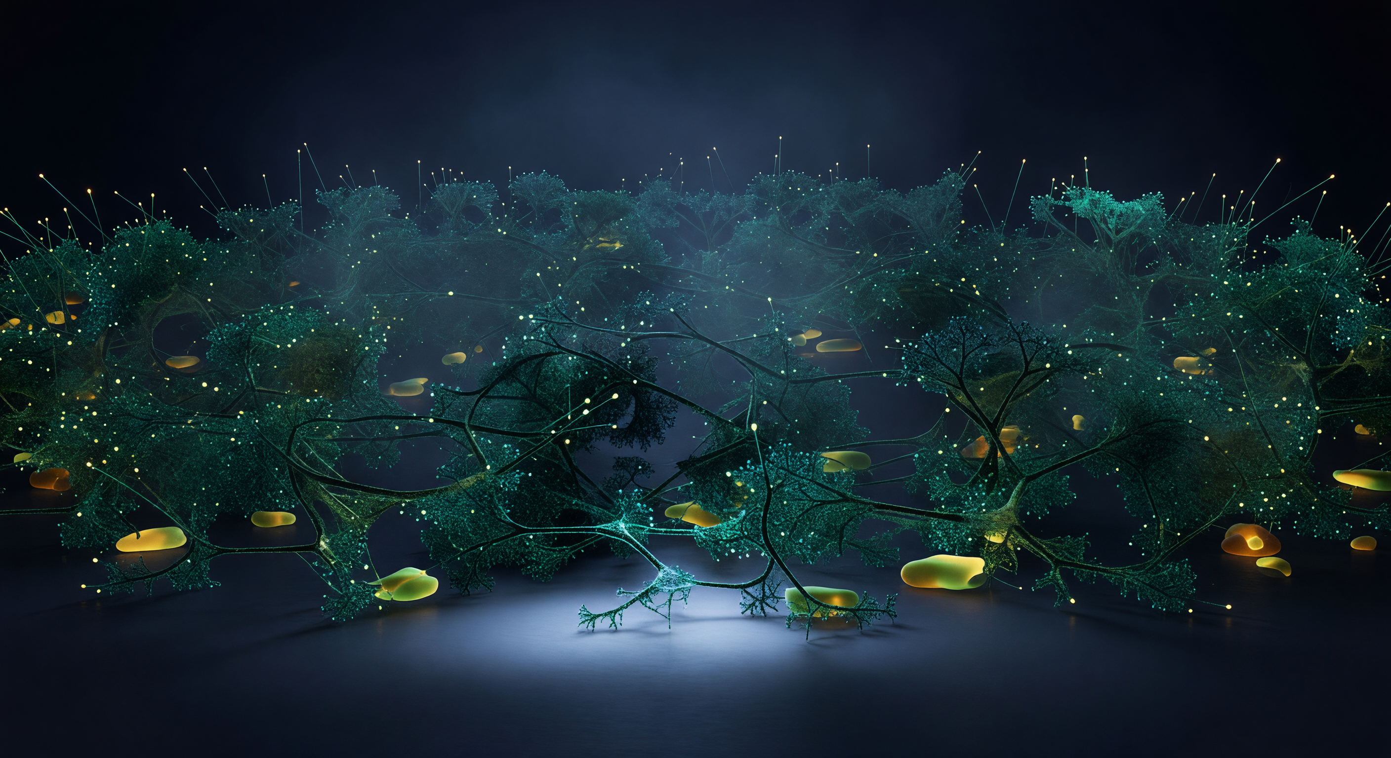
You stand at the outermost frontier of a living cell, suspended at the advancing tip of a lamellipodium where individual actin filaments — each just seven nanometers thick — branch from Arp2/3 complexes at precise seventy-degree angles, building a fractal canopy of electric teal and cold jade that radiates inward like an endlessly repeating coral reef frozen in the act of growing. Beneath you, the substrate is illuminated only by an evanescent wave penetrating a few hundred nanometers upward, a cold TIRF glow that catches the warm amber halos of focal adhesion plaques — molecular anchor complexes the size of boulders from this vantage, their integrin-rich surfaces bridging the extracellular matrix to the cytoskeletal machinery driving the whole cell forward. The actin network is not random but exquisitely regulated: Arp2/3 branching, capping proteins, and cofilin-mediated severing balance polymerization at the leading edge against disassembly further inward, generating the protrusive force that pushes membrane against resistance through nothing more than the chemistry of monomer addition. To either side, filopodial bundles — parallel actin arrays distinct from the branched lamellipodial mesh — project outward into the extracellular void like luminous antennae, their tips sensing substrate chemistry and topography in real time. Everything here trembles with thermal energy, macromolecular crowding thickens the medium to a dense gel, and the branching architecture continues inward layer behind layer until density alone swallows the light.
Eukaryotic cells (tissues)