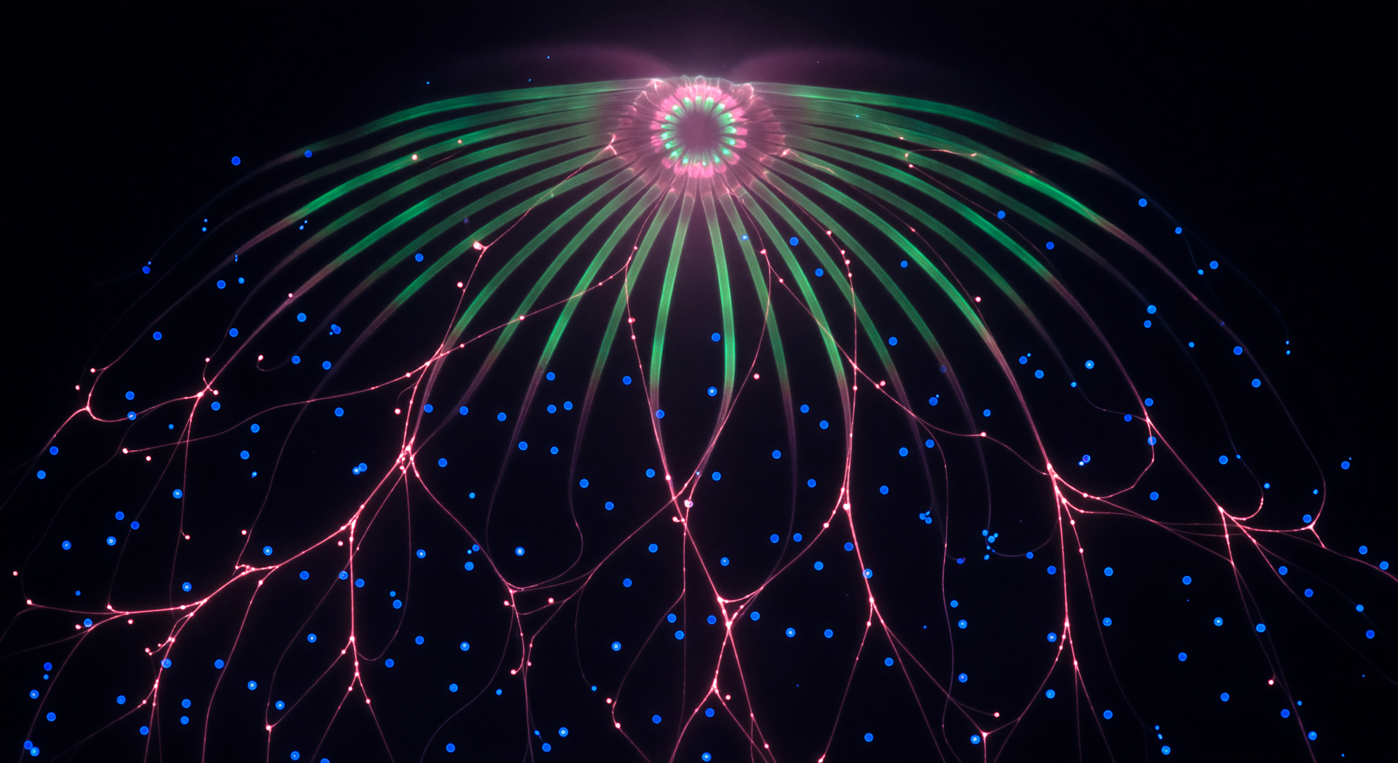
You are suspended inside a living animal smaller than a fingernail, yet the space you occupy reads as vast — the interior of a two-millimeter cydippid larva rendered in light-sheet fluorescence, its absolute black background interrupted by cold blue DAPI-labeled nuclei drifting through the mesoglea like sparse stars at varying depths, each a small hard cerulean sphere marking a cell nucleus within the organism's water-rich extracellular matrix. From that darkness, a non-hierarchical web of magenta filaments — FMRFamide-immunoreactive fibers of the diffuse nerve net, a neural architecture so ancient it predates bilateral symmetry — radiates in every direction without center or hierarchy, branching and re-branching asymmetrically, nodes glowing where fibers converge, casting soft rose halos into the void around them. Eight arcs of acid green sweep overhead like the ribs of a vaulted cathedral, each one marking a serotonin-positive comb row band — the sensory and locomotor infrastructure that coordinates the metachronal ciliary waves driving this animal through open water — their clean lateral edges imposing a spatial grammar on the otherwise anarchic filament web below. At the zenith, the aboral apical organ blazes as a near-white circular halo where magenta and green signals co-localize in the statocyst's lithocyte crown, the gravity-sensing structure that tells the animal which way is up in a featureless ocean — and from here, drifting through overlapping focal planes where nearer filaments are razor-edged and distant ones dissolve into glowing threads, the felt scale of this two-millimeter creature becomes indistinguishable from a nebula.
Other languages
- Français: Intérieur du réseau neural fluorescent
- Español: Interior de red neural fluorescente
- Português: Interior da rede neural fluorescente
- Deutsch: Fluoreszenz-Nervennetz Innenraum
- العربية: داخل الشبكة العصبية الفلورية
- हिन्दी: प्रतिदीप्ति तंत्रिका जाल अंदर
- 日本語: 蛍光神経網の内部
- 한국어: 형광 신경망 내부
- Italiano: Interno della rete neurale fluorescente
- Nederlands: Fluorescerend neuraal netwerk interieur