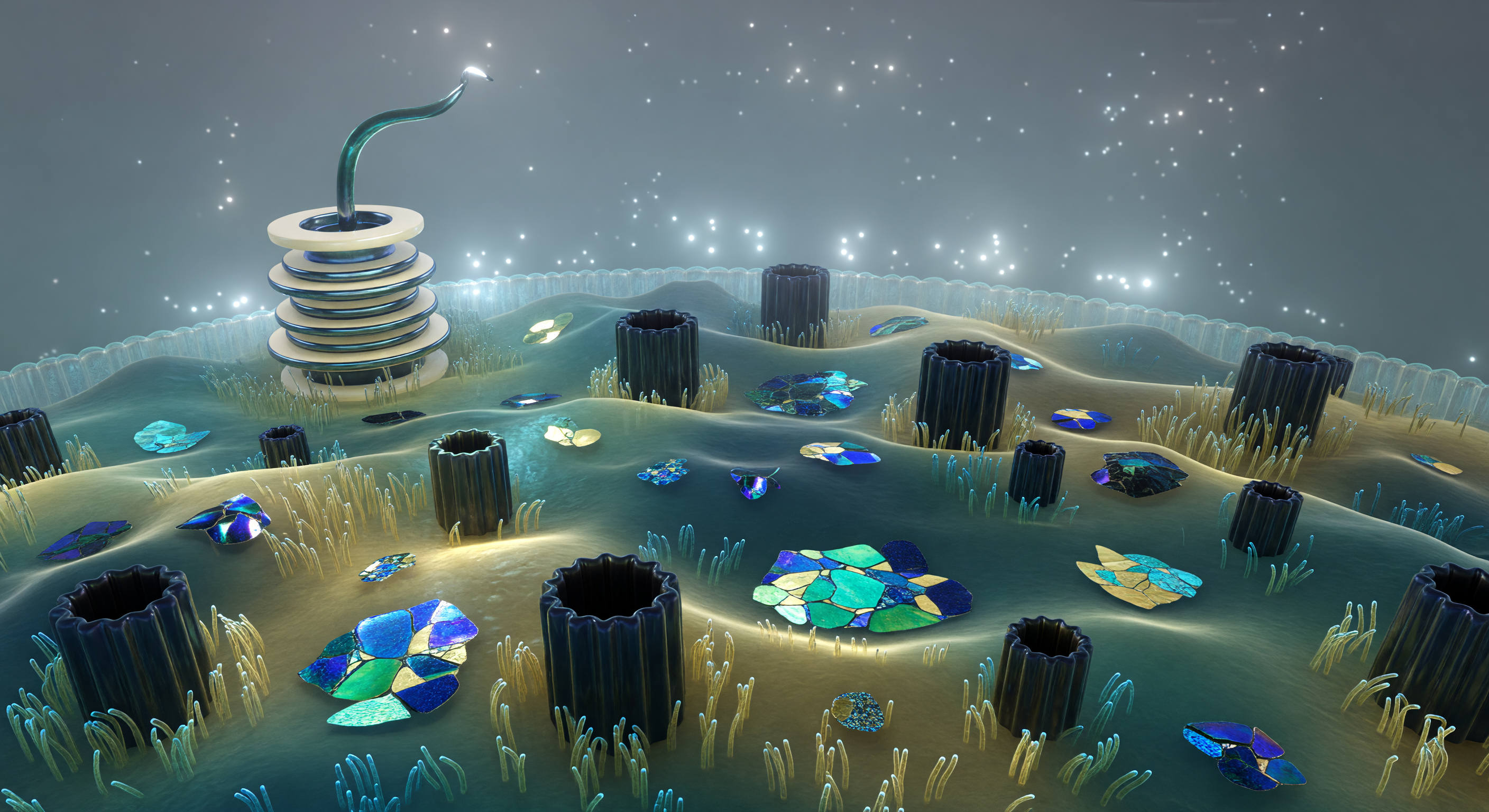
You hover over a living landscape that defies easy comprehension — the outer membrane of an *E. coli* cell, its lipopolysaccharide surface rolling in slow thermal waves driven by the constant agitation of molecular-scale energy, a bilayer so chemically asymmetric that its outer leaflet is armored with Lipid A and long polysaccharide chains rather than the phospholipids that line its inner face. Every twenty nanometers or so, OmpF and OmpC porin trimers pierce the membrane as rigid beta-barrel cylinders, their hollow interiors forming aqueous channels just wide enough to admit small solutes while excluding larger molecules, and their protein surfaces darkly iridescent where cold ambient light catches the geometric fluting of their rims. At this scale, Brownian motion is not a background abstraction but a felt reality — the entire plain shudders with thermal fluctuations that, at larger scales, would average to stillness, and the dense ionic atmosphere pressing against the membrane surface represents the Debye screening layer, a diffuse electrostatic haze a few nanometers thick where counterions crowd against the membrane's net negative charge. On the far horizon, the flagellar basal body rises as a multi-ringed protein machine spanning the full thickness of the cell envelope, its L-ring anchored in the outer membrane, its MS-ring embedded in the inner membrane far below, and its hook already curving upward into the viscous aqueous medium — the coupling interface for a rotary motor that, when running, spins at hundreds of revolutions per second and drives the cell through a fluid that, at this scale, behaves more like cold syrup than open water.
Other languages
- Français: Surface LPS Sous Lumière Froide
- Español: Superficie LPS Bajo Luz Fría
- Português: Superfície LPS Sob Luz Fria
- Deutsch: LPS Oberfläche Im Kryolicht
- العربية: سطح LPS تحت الضوء البارد
- हिन्दी: क्रायो-प्रकाश में LPS सतह
- 日本語: 極低温光のLPS表面
- 한국어: 저온 빛 아래 LPS 표면
- Italiano: Superficie LPS Sotto Luce Fredda
- Nederlands: LPS Oppervlak In Kryolicht