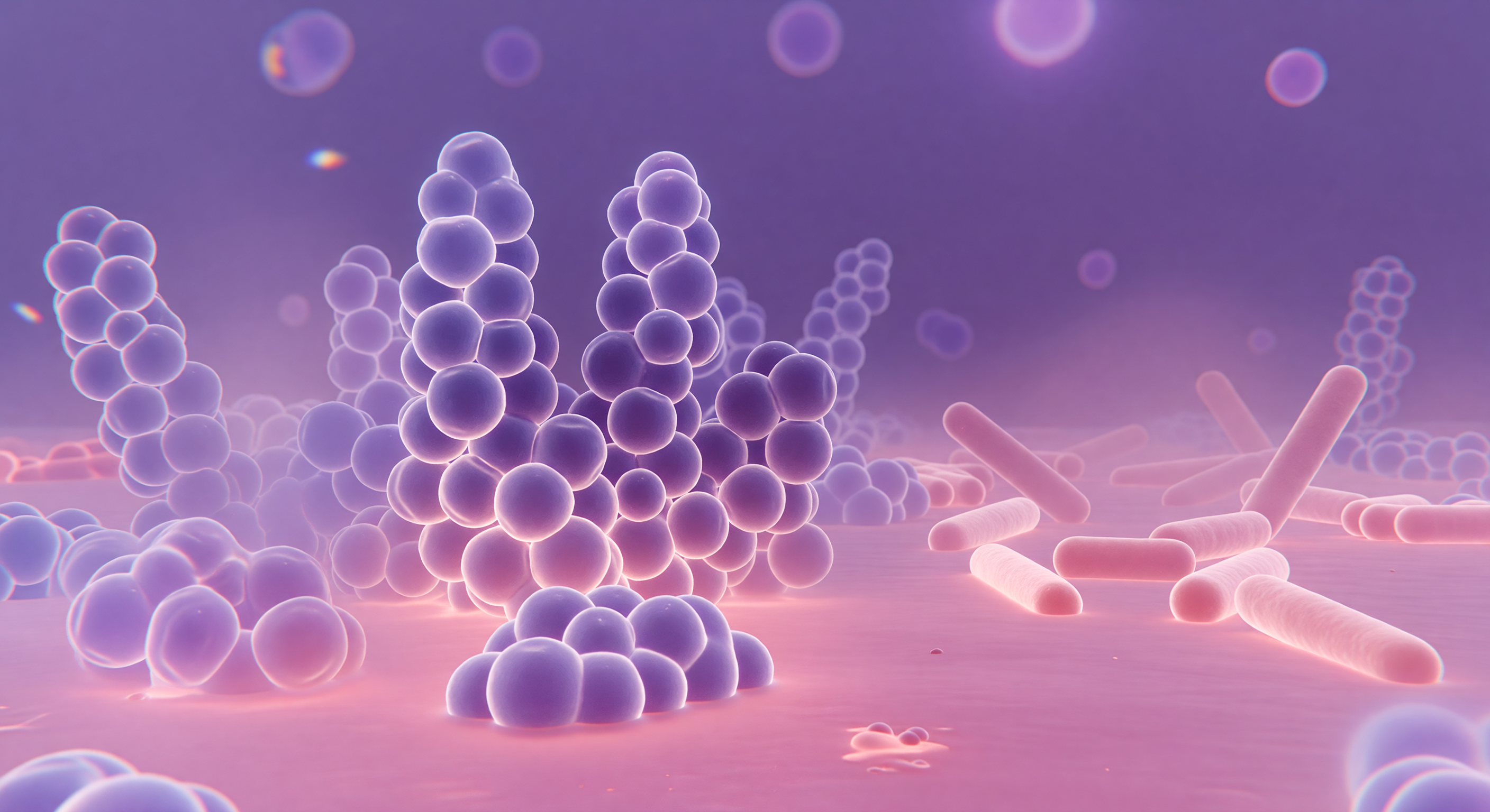
You stand inside a world saturated in deep violet and warm rose, surrounded by towering spherical masses that cluster overhead and on all sides like enormous soap bubbles compressed by their own abundance, each one a *Staphylococcus aureus* coccus whose thick Gram-positive peptidoglycan wall has greedily bound crystal violet dye, staining the entire landscape a bruised, luminous purple. The light arrives from everywhere at once, transmitted through the specimen like illumination through stained glass, casting no shadows but instead giving every curved surface an inner glow, while around each sphere's widest girth a faint diffraction corona shimmers — a physical consequence of visible light bending around objects barely larger than its own wavelength, ~400–700 nm meeting a 1-μm cell, the optics of glass and biology negotiating at their shared limit. Depth collapses abruptly: cells displaced even slightly above or below the focal plane dissolve into translucent lavender halos, their membranes bleeding into the warm safranin-pink counterstain that pools across the ground like rose-tinted water between the cluster's bases, marking the Gram-negative *E. coli* rods in the far quadrant — pale dusty pink, three times your length, their thinner peptidoglycan unable to retain the primary stain. At the visual periphery, chromatic fringes of blue and amber rim the ghost outlines of distant cells, honest artifacts of glass optics that make this single luminous focal plane feel like a window cut through an infinite layered city of living architecture extending upward and downward, purple and pink, far beyond sight.
Other languages
- Français: Monde de la Microscopie à Coloration de Gram
- Español: Mundo de Microscopía con Tinción de Gram
- Português: Mundo da Microscopia por Coloração de Gram
- Deutsch: Gramfärbung Lichtmikroskopie Welt
- العربية: عالم مجهر تلوين غرام
- हिन्दी: ग्राम स्टेन सूक्ष्मदर्शी जगत
- 日本語: グラム染色光学顕微鏡の世界
- 한국어: 그람 염색 광학 현미경 세계
- Italiano: Mondo della Microscopia a Colorazione di Gram
- Nederlands: Gramkleuring Lichtmicroscopie Wereld